Articles
Congenital Anomalies
Vol. 28 Supplement October 1988Proceedings of the Second Meeting of the International Federation of Teratology Societies
held on July 14-16, 1988
at Kyoto International Conference Hall, Kyoto, JapanSymposium II: Identification of human teratogens
Article below published online with the kind permission from Congenital Anomalies and authors. Many thanks to Dr. Kohji A. Matsui for the assistance.
Cong. Anom., 28 (Suppl.): S59-S69, 1988
HOW IT CAME ABOUT THE FINDING OF METHYL MERCURY POISONING IN MINAMATA DISTRICT
Yoshitaka HARADA and Kaneki NODA
Japanese Red Cross Kumamoto Red Cross Blood Center, Kumamoto 867, and Noda Clinic, Yabe-machi, Kumamoto 861-35, Japan
ABSTRACT
On April 21, 1956, 5-year-old girl who complained of various cerebral symptoms came to consult and check into Dr. Noda's pediatric clinic. It was on April 29 that her sister, a 3-year-old girl, came to the clinic under similar signs. He notified these two patients suffering from an unclarified disease with cerebral signs to Minamata Health Center on May 1. This was the start of the matter of Minamata disease. In consequence of an epidemiologic investigation by the Health Center, it became clear that a large number of patients like them appeared around the same area. In August, 1956, Medical Study Group of Minamata Disease was organized in Kumamoto University and started investigating the cause. In November, 1956, Medical Study Group considered the true symptom as a poisoning by heavy metal which was mixed into the sea foods of the spot. It was in July, 1959, that they finally confirmed this poison as mercury. Around 1958, we noticed that there were many patients with signs of so-called cerebral palsy who were born in the same area of Minamata disease and were born almost at the same time. As a result of investigation, we found that this cerebral palsy was congenital Minamata disease.
KEY WORDS
congenital Minamata disease, metyl mercury, cerebral palsy, fish, shellfish, cat
On April 21, 1956, a 5-year-old girl who complained of various cerebral symptoms came to consult and check into Dr. Noda's pediatric clinic (Harada, 1968a). He undertook various clinical examinations, but one week passed without a well-grounded diagnosis. It was on April 29 that her sister, a 3-year-old girl, came to the clinic under similar signs. Dr. Noda inquired her mother about detailed circumstances, and took notice of a possibility that there might be other patients in the neighboring family. Then Dr. Noda notified these two patients suffering from an unclarified disease with cerebral signs to Minamata Health Center on May 1. This was the start of the matter of Minamata disease (MD). In consequence of an epidemiologic investigation by Health Center, it became clear that a large number of patients like them appeared around the same area.
In August, 1956, Medical Study Group of MD was organized in Kumamoto University and started investigating the cause (Table 1). In November, 1956, Medical Study Group considered the true symptom as a poisoning by heavy metal which was mixed into the sea foods of the spot. And in July, 1959, they comfirmed this poison was a methyl-mercuric compound.
About 1958, we noticed that there were many patients with signs of so-called cerebral palsy (CP) who were born in the same area of MD and at the same time. As a result of investigation, we found that this CP was congenital MD.
PROGRESS OF THE STUDY OF MINAMATA DISEASE
Here, we will explain about the progress until we verified that the causative material of these symptoms was methyl mercury (Table 2).
Epidemiological special features at the beginning
- Almost all the patients lived in the same area of Minamata Bay.
- Disease mainly broke out among the fishermen and their family.
- It broke out in all ages except infant, without distinction of sex.
- The rate of patients was the same on both sexes in children, but higher in the male than the female in adults.
- There was no regular interval between the out-break of disease in a family, for example, several days in some cases, but several years in others.
- The death rate was high.
- Agricultural products and drinking water were not suspected as the cause.
- Every patient had eaten fish and shellfish from Minamata Bay.
- Many cats with the same signs as patients of MD were found in the same area and at the same time.
The clinical features
We could not find the specific changes in the family history and past history of these patients.
The main symptoms in adults (Tokuomi, 1968) were ataxia, concentric constriction of the bilateral visual field, sensory disturbance, auditory disturbance, extrapyramidal signs such as muscular rigidity and involuntary movement, and mental signs such as slight intellectual deterioration and marked emotional instability.
In infant cases (Harada, 1968a), there was no fever, and general condition was not so impaired at the beginning, while the disturbance in coordination appeared gradually. For example, use of chopsticks, tying shoestrings, and buttoning one's clothes were impaired. Following these, disturbance in gait and speech developed. Difficulty in mastication and swallowing, and blurring of vision were found in every case. Some patients complained of numbness of the mouth surroundings and the extremities, and pain in the joints and finger tips. In both severe and moderate cases, involuntary movements were noticed. In acute cases, tremor, clouded consciousness, convulsions and rigidity of the extremities were observed.
We could not reveal the special and pathological findings by routine clinical and laboratory examinations.
Post-mortem examination in 1956 (Tekeuchi, 1968)
The first autopsy of subchronic case was made on August 3, 1956, and the second autopsy of subacute case was performed on September 5, 1956. These results showed that MD consisted of a toxic encephalopathy without primary inflammation.
Preliminary report by Medical Study Group
In November, 1956, Study Group put their thought together that MD did not belong to infectious disease but to intoxicative disease, being caused by eating a large amount of fish and shellfish caught in Minamata Bay. As the noxious factor contaminating the fish and shellfish, several kinds of metals and metalloids, especially manganese, selenium and thallium were considered.
The first experimental study feeding the fish and shellfish to cats
At the same time, many cats which suffered from unsteady and slow movements, ataxic gait and paroxysmal convulsion were found in the same area of outbreak of MD. Autopsy finding of the brain of these cats bore striking resemblance to the feature of patients of MD. In the experimental study, the same clinicopathological features in cats fed fish and shellfish caught in Minamata Bay were seen as in cats sicked spontaneously in Minamata district. From these facts, we found that this disease was caused by eating fish and shellfish caught in Minamata Bay. But, it was not clear which metals or metalloids caused this disease at that time.
Then, we started the studies to determine the causative agent, for example manganese, selenium, thallium and the like. But the posonings of these metals have some difference in clinical, epidemiological and pathological findings (Table 3). So, we had not the same opinion of the causative agent about this disease.
In 1958, from the investigation of post-mortem examination of 10 cases and the literature available on this subject, we observed that this disease was very similar to the clinical features and pathological findings caused by alkyl mercury poisoning reported by Hunter and Russell (1954). Until that time, the poisoning of organic mercury, acute or chronic, was already reported in some cases clinically, but only one case took an autopsy.
Then the measurement of mercury content was begun by dithizone method. Mercury content of the brain, liver and kidney of autopsy cases increased slightly in chronic cases and extremely large in acute and subacute cases. Mercury was deposited more abundantly in the parenchymatous organs, such as kidney and liver, than in the brain.
These results were seen, similarly in the animals spontaneously sicked and experimentally fed fish and shellfish. And also, a large amount of mercury in the fish and shellfish caught in Minamata Bay was estimated.
On September 26, 1958 the council of the Study Group found the view that the organic mercury was most doubtful as noxious factor of this disease. At this stage, it was not clear what kind of organic mercury was concerned.
The investigation of reproducing similar features by taking the various kinds of organo-mercury compounds was begun. In 1961-62, Uchida et al. (1961) extracted a sample chemically from shellfish caught in Minamata Bay, and identified it as a methyl-methyl-mercuric sulfide (CH3-Hg-S-CH3).
Subsequently (1962-63), Kondo (1964) extracted it from Hormomya mutabilis and Venus japonica and identified it as methyl-mercuric chloride (CH3HgCl). Furthermore, Irukayama et al. (1962) extracted it from the slag of the chemical plant of the Minamata Factory and identified it as CH3HgCl and this made clear the process of formation of organic mercury.
In the experiments of using these substances to cats and rats, it was found that the same symptoms and pathological features as MD were produced. Now, it is thought that the agent which brings the pathological changes of MD is methyl mercury group (CH3Hg-).
CONGENITAL MINAMATA DISEASE
Next, we will explain about the congenital MD (Table 4). When we investigated the epidemiology of MD in 1956-57, we took notice of the fact that there were may patients who suffered from CP in the same area. So, we started the investigation about these CP.
The characteristic features of epidemiological results (Nagano et al., 1960; Harada, 1968b)
- The fetal period of many cases coincided with an out-break of MD.
- The regional distribution of out-break was the same.
- Many families of patients who suffered from CP had one or more patients of MD.
- Many families were engaged in fishing.
- The rate of out-break of CP in this district was high.
- No evidence of hereditary illness
- No abnormalities in delivery history
- There was no maternal abnormality during pregnancy and the mothers had normal delivery.
Clinical features (Nagano et al., 1960; Harada, 1968b; Harada and Moriyama, 1976)
Their abnormality was discovered by delay of development of motility. Mastication, swallowing and self-feeding was impaired. Their body and psychological development was severely impaired. There was no malformation.
Their PEG showed several abnormal features. Abnormal EEG was found in many patients. These patients had normal hormonal function and no chromosomal aberration. Mercury content in the hair of almost all the patients and their mothers was higher than that of normal children and their mothers in the same district (Table 5). A correlation between the mercury content in the hair of mothers and that of their children was found. The mercury content in umbilical blood of the babies born in Minamata City was slightly higher than those of other districts. It seems that organic mercury entered into the fetuses from their mother's blood through the placenta.
From the epidemiological and clinical features, we suspected that the causative agent of these CP was the same as that of infant and adult MD. This supposition was made sure by the autopsy finding and experimental study treated with the methyl mercury to the pregnant cats.
Post-mortem examination (Takeuchi, 1968)
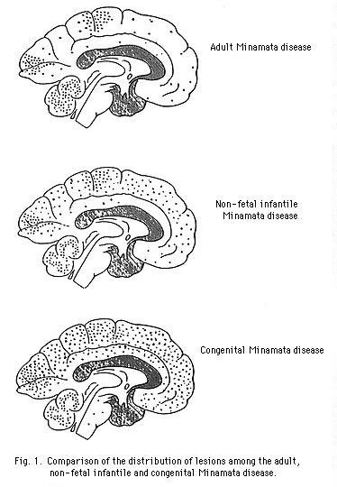
Two patients (3-year-old girl and 6-year-old girl) died in 1961 and 1962, respectively, and came into the autopsy. Post-mortem examination revealed that there were significant changes in the central nervous system, but other organs were never or only slightly affected. These patients had two characteristic pathological findings, one of which coincides with the neuropathological features seen in infantile MD, and the other is related to the age at fetal exposure. The latter is represented by the hypoplastic and dysplastic findings in the central nervous system (Table 6).
The sererity of the lesions and the extent of the involvement differed with age (Fig. 1). There was a tendency that the younger the patient was, the more severe the symptoms were, and the more wider the lesions were. Commonly, it seemed that an immature brain was more easily impaired than a mature one.
In the post-mortem examination, it was revealed that mercury content of the brain, liver and kidney was larger than that of normals. This offered an additional evidence in the diagnosis of mercury poisonings in these cases.
Animal experiments
At the beginning, namely, when we found out many CP in this district, we thought that these congenital CP had a close relationship to MD. While organic mercury was considered as the causative agent of MD (Nagano et al., 1960), we set to work for finding experimentally the effect of mercury on the pregnancy and fetus of animals: 1. the course of pregnancy, 2. fetal development, 3. mercury content in some organs of the mother and fetus, and 4. microscopic and electron-microscopic features of the mother and their offering (Harada and Moriyama, 1976).
The mercury content of the hair, brain, liver and kidney markedly increased in both dams and young. The brain findings resembled to those of cats spontaneously illed near Minamata or those fed fish or shellfish experimentally.
We diagnosed these CP as congenital MD from 1. epidemiological investigation, 2. clinical signs and laboratory findings, 3. post-mortem examination, and 4. experimental studies.
CONCLUSION
- MD is a poisoning of methyl mercury which is seen in people who ate fish and shellfish contaminated by methyl mercury. In this case, the brain is a main part of damage.
- In case of pregnant woman, methyl mercury intrudes into fetus from mother through the placenta, and causes the congenital MD.
- The brain damage of congenital MD is more extensive and severe as compared with that of the infant or adult. Therefore, clinical features are more serious in congenital cases.
On Reality. Publisher and editor: Bo Walhjalt. ISSN 1650-9323.
© Bo Walhjalt and authors. | Comments on this page
Latest update 2002-12-05